Computational Tools and Software
Analyzing cellular morphodynamics and biosensor activity
Protrusion Dynamics
Protrusions play a crucial role in many physiological processes including cell migration during development, wound healing and immune response. However, quantification of highly dynamic protrusion across a broad range of shapes and sizes creates a serious challenge for both imaging and statistical analysis. Here we developed a powerful computational platform CellGeo that makes precise definition and identification of protrusions possible and allows automated tracking and analysis of protrusion dynamics to be unbiased, labor efficient, and comprehensive. [Published here: J Cell Biol. 2014 Feb 3; 204(3): 443-60. doi: 10.1083/jcb.201306067. [ncbi pmc]
Filopodia identification and tracking
Thin protrusions like filopodia are often hard to visualize with sufficient spatiotemporal resolution and a high signal to noise ratio. Kymograph (line scan) approaches do not work as well for cells with complex geometries and especially curving, highly dynamic filopodia. Even if the tips of filopodia are identified, it is hard to define and consistently measure filopodia width and length in an automated manner. It is also hard to define a generic distance measure for tracking multiple closely located filopodia that grow out and retract at different time points.
MovTresh module of the CellGeo software package not only automatically thresholds every frame of the movie and highlights cell outline, but also provides a convenient interactive controls to adjust threshold values and outline filopodia as accurate as the signal-to-noise ratio allows. BisectoGraph module maps an arbitrarily complex shape onto a tree-graph that allows clear definition and identification of filopodia tips, bases and central lines automatically and consistently across all geometries and sizes. FiloTrack module provides interactive controls for adjusting tracking parameters and measuring filopodia lifetime, number and length. Each tracked filopodia can be individually visualized for easy assessment of the accuracy of the results.
Broad protrusion activity
When both broad protrusions and filopodia present at the cell edge, visual perception of their individual contributions to the cell dynamics is obscured. The total change of cell area is not always an accurate indicator of protrusive or refractive activity, because the cell can undergo significant but randomly distributed protrusions and retractions with little change of the total area.
The tree-graph representation of the cell outline used in the BisectoGraph module of the CellGeo software package is a perfect tool for segmenting out all filopodia and extracting the underlying cell body for further analysis. This way, thin and broad protrusions of the same cell can be analyzed simultaneously but independently of each other. ProActive module visualizes protruding and retracting parts of the cells (in different colors) and quantifies protrusive and refractive activity as a function of time using both the area and velocity measures.
Growth cone dynamics in neurons
Once again, the tree-graph representation of the shapes used in ConeTrack module of the CellGeo software package provides a way to automatically extract and track outlines of each growth cone with just two user-specified criteria.

Cellular morphodynamics
Before reaching a final state after drug treatment, cells can undergo transient morphology transformations that are significantly different from the final shape or motion type. Quantitative analysis of such transient dynamics is particularly important in dissecting functional roles of homologous cytoskeleton regulators. However, this requires a tool that is capable to classify cell motion types as the time goes.
By introducing a set of parameters that characterize major aspects of shape changes, such as the rate of area change and polarization factor, cell motion can be represented as a trajectory in parameter space with distinct regions that correspond to different types of motion. Based on this approach, our GUI SquigglyMorph classifies cell motion as uniform spreading, uniform shrinking, polarized spreading, polarized motion, or polarized shrinking at each time frame of the imported movie. Published here: Methods Cell Biol. 2014;123:409-27. doi: 10.1016/B978-0-12- 420138-5.00022-7.
Spatiotemporal distribution of GTPases at the cell edge
To establish correlations between the activity of a GTPase and the cell edge motion, spatiotemporal distribution of active GTPase needs to be visualized and quantify with respect to the cell edge and its velocity. However, it is important that such quantification is automated and includes the whole (or most of the) cell edge to avoid selection bias and poor statistics.
The GUI LineScan automatically builds scan lines of a user-defined length perpendicular to the cell edge along the whole cell outline. To calculate edge velocity, LineScan utilizes an elastic model to find the optimal correspondence between boundary points at the consecutive time frames. Finally, LineScan displays and saves biosensor activity profile as a function of edge velocity and the distance from the edge for individual time frames, but also for the whole movie by averaging activity profiles over time. Published here: Methods. 2014 Mar 15;66(2):162-7. doi:10.1016/j.ymeth.2013.08.025.
Polarization factors in yeast
When using multiple fluorochromes for tracking proteins of interest and, simultaneously, for detection of individual cell in culture with membrane and nuclei markers is not an option for technical reasons, quantification of the proteins distribution in individual cells become a problem. Automated cell segmentation is still possible, but it has to be built based on other hints, such as an a priori expected shape of the cells.
The GUI SegmentMe is a geometry-based segmentation tool, which allows segmentation and tracking of yeast cells in tight clusters. In addition, SegmentMe provides a set of quantitative measures, such as size, velocity, MSD, etc., for the analysis of protein clusters (spots) within segmented cells in both 2D and 3D data sets. Published here: Methods Cell Biol. 2014;123:409-27. doi: 10.1016/B978-0-12- 420138-5.00022-7.
THE software collaboration (Tsygankov Hahn Elston)
Software resulting from a collaboration of the Tsygankov, Hahn, and Elston Labs.
1. CellGeo (multi-module platform for cell protrusion analysis: automated identification and tracking of protrusions of different scales and with arbitrarily complex geometry).
2. LineScan (automated full-edge line-scan for quantification of correlations between intracellular fluorescence intensity and cell edge velocity).
3. SquigglyMorph (automated classification of cellular morphodynamics)
4. SegmentMe (automated segmentation and tracking of tightly packed cells with simple geometry; allows quantification of intracellular clustering.)
5. EdgeProps (A method for correlative analysis of signal activity, edge velocity, protrusion persistence and orientation; the approach is not sensitive to roughness of the cell edge, i.e. overcomes the limitations of LineScan and Gaudenz Box methods; includes full-edge kymograph and other representations of the edge dynamic.)
Stochastic models of cell migration
Cell migration requires precise spatiotemporal regulation of the actin cytoskeleton. Key regulators of cell movement are the Rho family of GTPases. To investigate the role of Rho GTPases in cell migration, we developing stochastic models of cell movement to analyze time series data for the position of migrating cells. Our approach allows parameters that quantitatively characterize cell movement to be efficiently estimated from experimental data. Our preliminary results indicate that randomly migrating cells stochastically transition between distinct states of migration characterized by differences in cell speed and persistence. (Investigators: Richard Allen, Chris Welch, Klaus Hahn and Tim Elston).


Stochastic modeling of biochemical networks
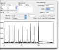
 We are interested in developing computational tools for studying stochastic effects in signaling pathways and gene expression. With David Adalsteinsson (Applied Mathematics, UNC), we have developed the software package Biochemical Network Stochastic Simulator (BioNetS) for efficiently and accurately simulating stochastic models of biochemical networks. BioNetS has a graphical user interface that allows models to be entered in a straightforward manner, and allows the user to specify the type of random variable (discrete or continuous) for each chemical species in the network. The discrete variable are simulated using an optimized implementation of the Gillespie algorithm. For the continous random variables, BioNetS constructs and numerically solves the appropriate chemical Langevin equations. The software package has been designed to scale efficiently with network size, thereby allowing large systems to be studied.
In collaboration with David Adalsteinsson and David McMillen
Adalsteinsson D, McMillen D, Elston TC. Biochemical Network Stochastic Simulator (BioNetS): software for stochastic modeling of biochemical networks. BMC Bioinformatics. 2004 Mar 8; 5:24
(Pubmed | Journal)


